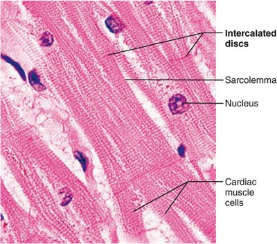
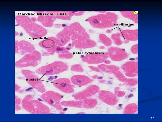
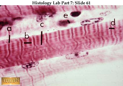
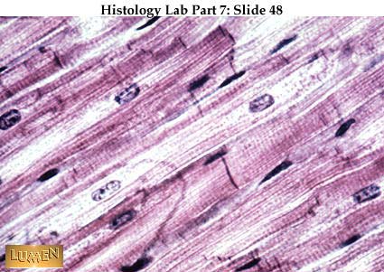
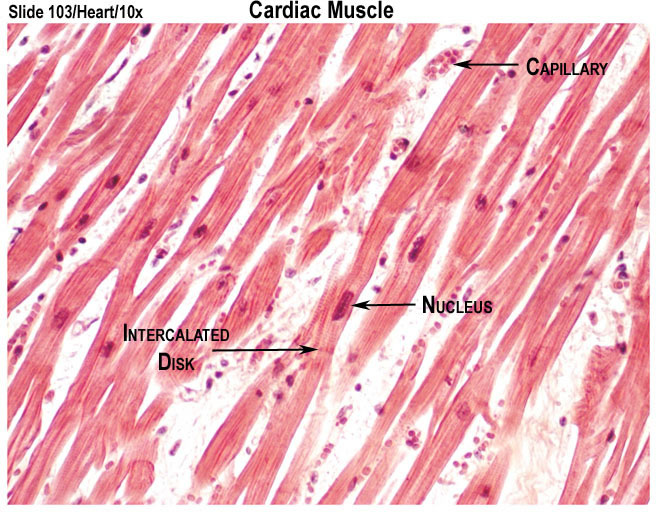
Cardiac Muscle Labeled Slide
In the sliding filament model myosin filaments slide along actin filaments to shorten or lengthen the muscle fiber for contraction and relaxation.
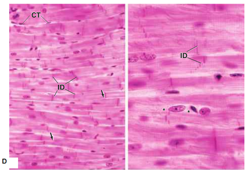
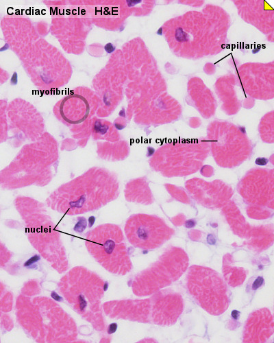
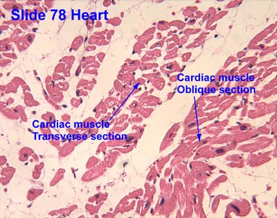
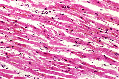
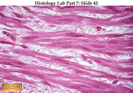
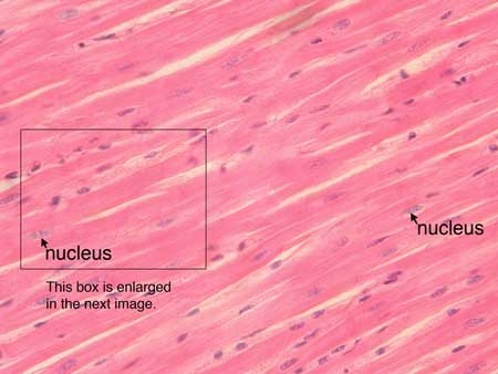
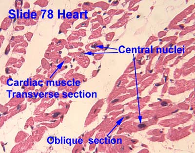
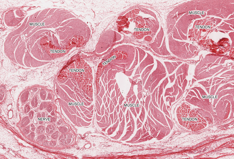
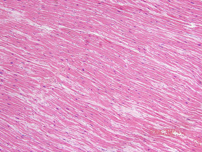
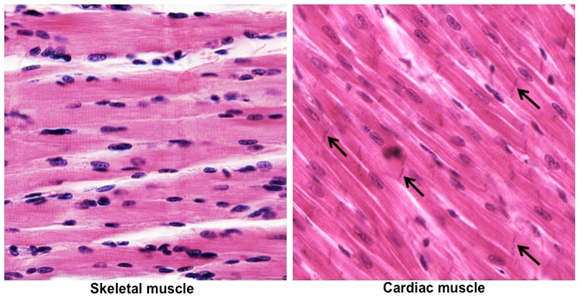
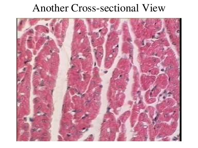
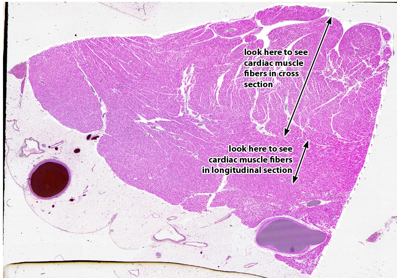
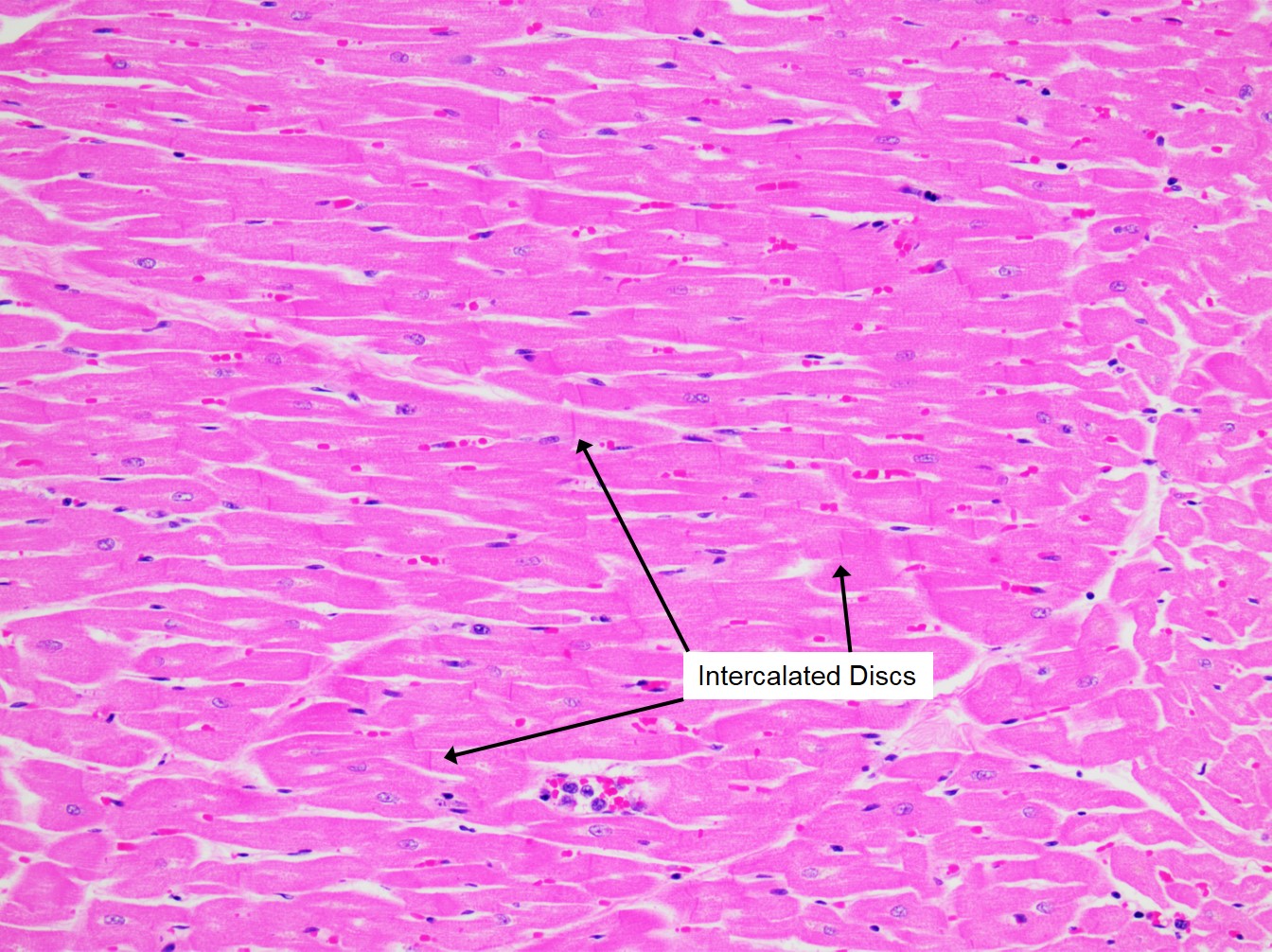
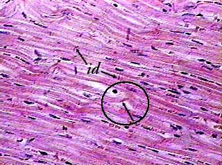
Cardiac muscle labeled slide. On any slide of cardiac muscleyou will see cells that have been sectioned in every possible directionfrom transverse to oblique to longitudinal. 305 heart ventricle he webscope note. The cells and their detailedstructure is best seen on cells that are sectioned longitudinally. This slide not in glass slide collection cardiac muscle will be studied in the wall of the ventricle of the heart.
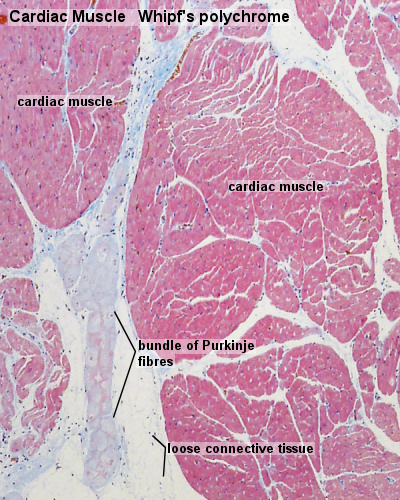
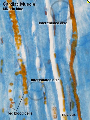
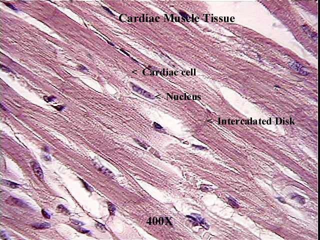
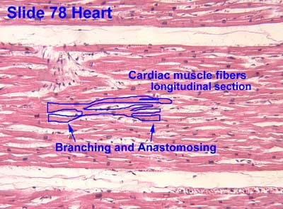
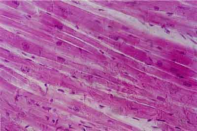
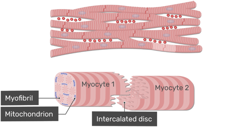
Connect to one another at intercalated discs. The individual cardiac muscle cells are arranged in bundles that form aspiral pattern in the wall of the heart. Mohammed abdul hannan hazari assistant professor department of physiology deccan college of medical sciences hyderabad 2. Cardiac muscle cells excitation is mediated by rythmically active modified.
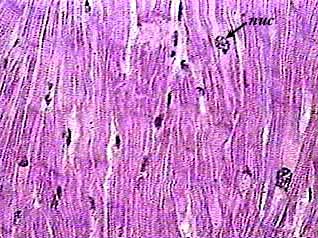
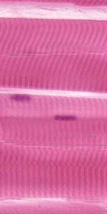
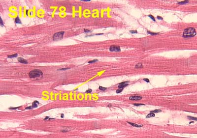
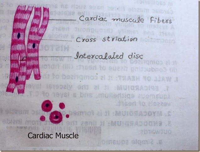
Cardiac muscle tissue works to keep your heart pumping through involuntary movements. Coronary arteries anterior view posterior view 4. The actual mechanical contraction response in cardiac muscle occurs via the sliding filament model of contraction. Similar to skeletal muscle cardiac muscle is striated and organized into sarcomeres possessing the same banding organization as skeletal muscle.
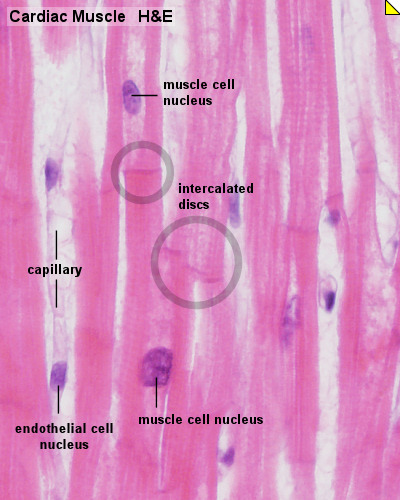
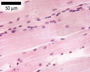
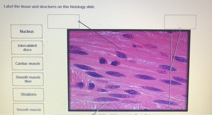
In comparison with skeletal muscle note the following differences. This is one feature that differentiates it from skeletal muscle tissue which you can control. Nuclei are oval rather pale and located centrally in the muscle cell which is 10 15 um wide. Heart location in the chest precordium 5.
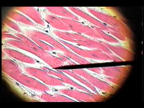
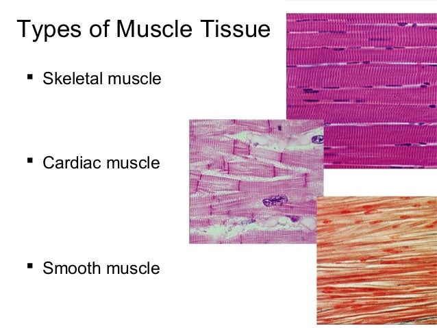
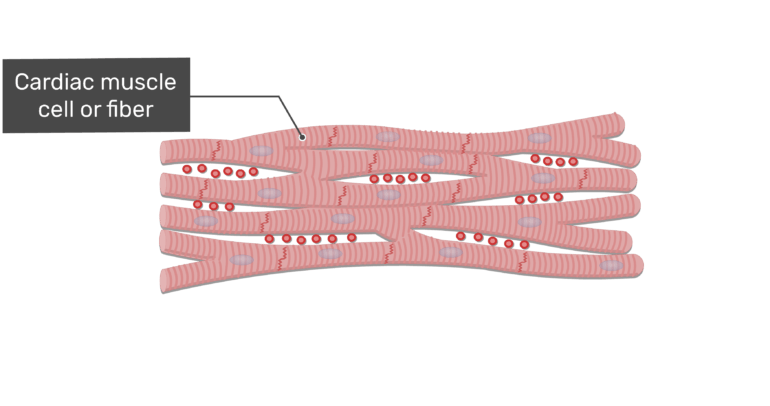
The pathway of contraction can be described in five steps. Cardiac muscle cells branch and form a three dimensional network. Cardiac muscle is striated involuntary muscle found in the heart wall. Histology of cardiac muscle 1 by.
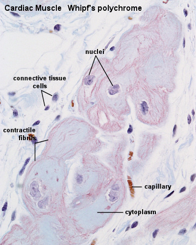
Cardiac muscle cells have rounded cross sections less than 25 um in diameter with a centrally located nucleus. Cardiac muscle and heart function cardiac muscle fibers are striated sarcomere is the functional unit fibers are branched. Introduction cardiac muscle the myocardium consists of cross striated muscle cells cardiomyocytes with one centrally placed nucleus. Cardiac muscle contains sarcomeres which in themselves contain myofibril proteins as well as actin and myosin filaments.
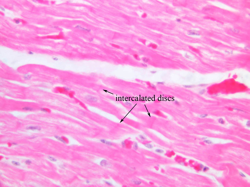
Cardiac contractile cells have intercalated discs that permit ions to pass between the cells which transmits the electrical impulse rapidly. However cardiac muscle fibers are shorter than skeletal muscle fibers and usually contain only one nucleus which is located in the central region of the cell.
Cardiacmuscleimages Main Medical Histology

Cardiac Muscle Images Stock Photos Vectors Shutterstock
Cardiac Muscle Histology Embryology

Blue Histology Muscle

Chapter 7 Page 8 Histologyolm 4 0


_040_02.jpg)


_100_04.jpg)